MX16 multi-slice computed tomograph from Philips
Orthopaedic Radiology
In 2007, the cooperation between the Orthopedic University Hospital and our institute was closed. Through this close integration of the different areas Radiological a comprehensive and consistent quality is ensured.
Since is the Department of Radiology at the Orthopedic University Hospital of the Technical supervision of Prof. Dr. med. Thomas Vogl Provided.
OA Dr. med. Matthias Heller is a Senior Physician in the Department
Range of services / equipment
Computed tomography
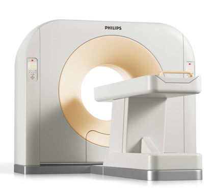
Philips Healthcare's state-of-the-art MX16 multi-slice computed tomography scanner allows us to offer our patients and referring physicians faster and more accurate diagnosis of diseases and accidental injuries.
Images in seconds
The CT scanner simultaneously acquires 16 cross-sectional images of the body region to be examined. This capability is based on Philips' proprietary technology that produces complete images of organs and vessels with the tightest slice staggering in a matter of seconds.
Lower radiation dose, higher image quality
The acquisition and assimilation of complex multi-layer images is made possible by a special integrated circuit developed by Philips within the detector group of the Mx16, which operates at speeds in the gigabit-per-second range. Analog detector signals are converted directly into a stream of digital data, significantly reducing noise. This, in turn, reduces the necessary radiation dose to the patient while increasing image quality.
X-ray
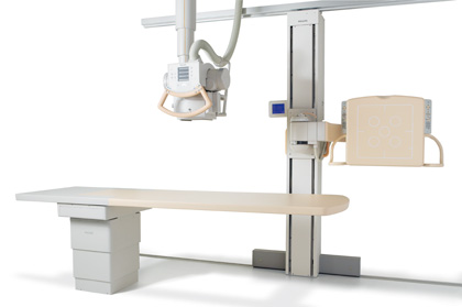
DigitalDiagnost VM Compact von Philips
Progress in orthopedic diagnostics
The DigitalDiagnost VM Compact from Philips is a single-detector stand that can be used universally for frontal, lateral and angulated images. It is suitable for all thoracic and skeletal diagnostics, as well as for imaging in trauma diagnostics.
The detector unit is equipped with a digital flat panel detector, which has a large image field of 43 cm x 43 cm, an image matrix of 9 million pixels and a digitization of the image with over 16 0000 gray levels corresponding to 14 bits.
The special advantage of this examination device is the attachment of the detector to a rotatable and tiltable C-arm stand.
Magnetic resonance imaging
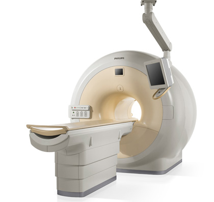
High-performance MRI Achieva 1.5T Nova von Philips
Our new high-performance magnetic resonance tomograph Achieva 1.5T Nova from Philips now offers our patients a significantly expanded examination spectrum. The magnetic resonance tomograph works without X-rays and thus avoids additional exposure of the patient to radiation. The device is extremely patient-friendly while providing high-resolution and high-contrast cross-sectional images of the human body.
The focus is on studies of the representation of:
- Orthopedic joint diagnostics
- Central and peripheral vascular imaging
- Cardiac diagnostics
- Head imaging
- Abdominal imaging
Team
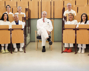
Responsible senior physician
Kontakt
Dr. med. Matthias Heller
069 6301-94252
069 6301-94323
Head MTRA
Kontakt
Heidrun Boller-Liedtke
069 6301-941951
069 6301-94323
