Microwave ablation
Microwave tumour ablation is a minimally invasive treatment method for primary and secondary tumours. Microwave ablation destroys the tumour from the inside by means of heat. At our institute, microwave ablation is used to combat tumours in the lungs and liver, among others. A special microwave probe is inserted into the tumour under CT control. A generator connected to the probe produces electromagnetic oscillations. The vibrations stimulate the hydrogen molecules in the tumours. The friction eventually generates heat that cooks the tumour from the inside.
What are the advantages of microwave ablation?
With this therapy it is possible to preserve more functional lung tissue in patients with lung tumors as can be saved by surgical resection. Hence this improves the life quality of the patient in cases of compromised lung capacity.
Performance of microwave ablation
In most of the cases the radiologist chooses the more patient friendly percutaneous entry which means a needle is inserted through the skin at the level where the therapy is to be done. The advantage of this method is that it is associated with little complications and that only a local anaesthesia is required.
After administration of local anaesthesia a small incision of about 2 mm is made. Simultaneously the patient is given a light sedative intravenously. Under CT-guidance the needle is slowly pushed forward to the region of interest which is then followed by radiofrequency ablation; heating of tumor cells. In case of tumor recurrence the radiofrequency ablation can be repeated.
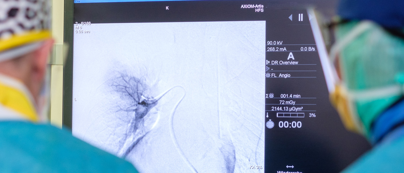
Information video: "How a microwave ablation works"
Information video in English
How is the success of the treatment checked?
A regular follow-up is done in form of CT or MRI examinations every three months to make sure that the whole tumor tissue has been destroyed. In this process reemergence of new tumors can be detected and treated again.
What complications can arise?
Different types of complications can arise depending on the type, size and localisation of the tumor. There is the possibility of bouts of cough, bleeding or the so called pneumothorax where air collects in the space between the lung and chest wall. Normally there is negative pressure in this space and results in compression of the lung when there is change in this pressure which must be reversed using a special catheter so that the lung is normally aerated. In such cases the patients must remain in the hospital for observation.
- Post-ablation bleeding can occur after an ablation. Since the radiologist constantly monitors the microwave ablation, he can intervene in time.
- Pneumothorax: Air enters between the lung and the pleural space and prevents the lung from expanding.
- It can happen that the treated tissue becomes inflamed and an abscess forms.
- Fever
- Local pain.
Are there any limitations to this type of therapy?
The possibility of application of this therapy depends on the tumor size. Because of the technical restriction it is not possible to treat tumor of very big sizes. On the other hand the tumors must have a certain size because it is not possible to sufficiently treat the smallest of tumors at microscopic levels. This means that there is a possibility of tumor recurrence.
Medtronic Microwave Ablation