Diagnostics in Magnetic Resonance Imaging
We offer the complete spectrum of magnetic resonance imaging. You will find more detailed information on some of the MRI examinations below.
Magnet resonance imaging allows one to produce cross-sectional images of human body without the use of radiation. Radiowaves produced within a spacious magnetic tube causes reactions within the body which is received as signal by coils placed on the examining region. These signals are converted into images by computer.
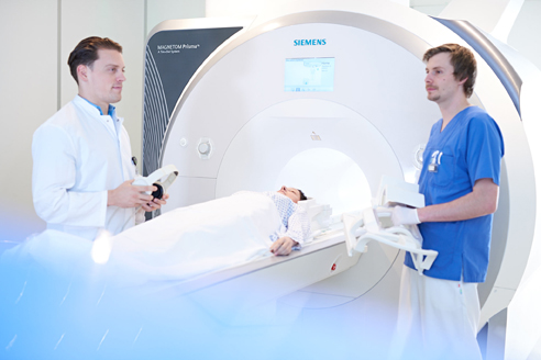
An MRI examination usually takes about 20 to 40 minutes. The patient is placed supine in a 60 cm broad (diameter) and 160 long tube. In cases of claustrophobic patients examination can be performed under mild sedation.
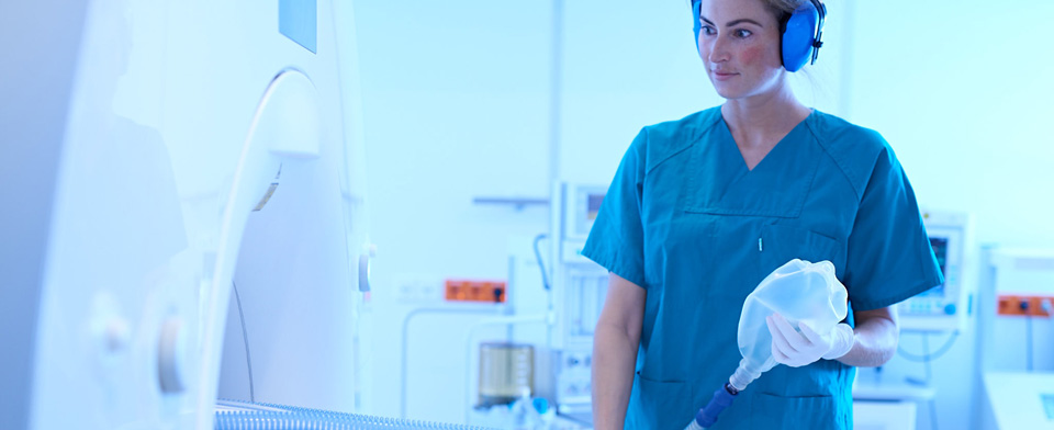
During the examination the patient hears loud banging-type of noise which is produced by activation of electromagnet (Radiofrequency) and unfortunately cannot be avoided. To dampen the noise the patients are provided with a head phone or ear plugs. It is very important that the patient lies still during the examination. Even minimal movements can lead to decrease in image quality. For examinations of the abdomen and thorax one receives breathing commands and must hold breath for short periods (10 to 20 seconds). Majority of examinations are performed with a special MRI-contrast medial (gadolinium) in order to better differentiate the soft tissues.
For the examination it is also important that the patient removes all metal elements from his/her body (e.g. spectacles, watch, jeweleries, hearing aids, tooth-implantations etc.). Also small zip or clip in brassiere can lead to disturbances in the image.
Important: if you have a cardiac pace maker you should not in any case enter the examination room!
Procedure and preparation before an MRI examination
An MRI examination usually takes between 20 and 40 minutes. You will be taken into a 60 cm (diameter) and 160 cm long tube while lying down. If you suffer from claustrophobia, you can be given a sedative beforehand.
During the examination you will hear loud knocking noises. These noises come from fast-switching electromagnets and unfortunately cannot be avoided. You will be given headphones or earplugs to protect you.
You must lie still during the examination. Even small movements can disturb the images. For examinations of the abdomen and chest area, you may have to hold your breath for a short time (10-20 seconds).
For the examination it is important that you take off all metal (e.g. glasses, watch, jewellery). Small zippers and bra straps can also interfere with the image. If you have a cardiac pacemaker, you must not use an MRI scanner under any circumstances!
Contrast agents considerably improve the significance of an MRI and can even reveal decisive information. Contrast media containing gadolinium are mainly excreted through the kidneys.
We only use contrast media after a comprehensive patient discussion and after careful consideration of whether the use of a contrast medium really offers relevant additional information. Of course, we administer the smallest possible amount. Our team only uses contrast media that have been tested according to international and German guidelines and are approved as medicinal products.
For more information on the subject of contrast media, read the patient information of the German Radiological Society (DRG) and the Professional Association of German Radiologists (BDR): https://www.drg.de/de-DE/3994/mrt-kt.
What you should bring to the examination
Reports and images of previous examinations are very helpful for the planning and evaluation of magnetic resonance imaging.
Please bring these with you if they were not performed at our Clinic.
For an MRI examination, you should bring current laboratory results with you that provide information about your kidney function:
- Creatinine value
- Glomerular filtration rate (GFR)
- TSH (thyroid).
These should not be older than 14 days. For this purpose, you can have your blood drawn by your referring doctor or family doctor. Please bring the results of the laboratory test with you.
Responsible senior physician Prof. Dr. med. Simon Martin