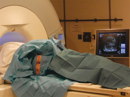
The Particularly large opening of the Puncture-MRT's allows the targeted sample collection under MRI Control.
MR-guided Biopsy of the prostate
The improved detection of PCa a growing number of patients required a precise histological classification the planning of further therapy. With the gold standard, the aforementioned transrectal needle biopsy of the prostate tumors can be detected systematic. However, a large number of biopsies is negative, so that further biopsies for adequate diagnosis is necessary. A targeted biopsy of prostate tissue tumorverdächtigem is not possible using the schematic biopsy. The precise puncture suspicious lesions under MRI control aims at filling this gap and provide a reliable result.

Indications for the Implementation of a Prostate with MRI Spectroscopy:
- Suspect Digital Palpation
- Elevated PSA Serum Concentration
- Screening
- Negative Punch Biopsies
Indications for performing an MRI guided Biopsy:
- Negative Punch Biopsies
- Small, only be visualized by MRI and MRS Tumor suspicious Lesions
Advantages of Prostate MRI and targeted Biopsy under MRI Control:
- Detection of even small suspicious Lesions in the Prostate.
- Tissue Analysis of diseased and healthy tissue.
- Avoid further negative Biopsies in MRI
- Targeted sample recovery from the Lesions.
- The healthy tissue can be preserved targeted compared against damage.
- Implementation as an Outpatient Treatment.
What must I bring?
For Performing an MRI-Guided Biopsy of the Prostate, you should bring the following laboratory values:
- Total PSA
- Ideally, the Course, showing the Date
- Ratio of free and bound PSA
The Results of preliminary Investigations (eg ultrasound, Biopsy-, medical reports) are very helpful in the Evaluation and Assessment of an MRI-Scan. We therefore ask you to bring us these findings in copy.
Expirationof the Investigation: Do I have to prepare for the exam?
For the Examination, you will 60 minutes Examination Time Schedule. You should shortly before the Examination to the toilet bowel and bladder emptying, so that the bowel is not too much later filled with the Puncture. Please be fasted prior to puncture. Generally MR Examinations can not be performed in patients with Pacemakers. Do you use Artificial Heart Valves or Inner Ear Prostheses have been implanted, must be clarified before the Investigation, whether they are MRI-Compatible. To do this we need your Implant-ID Card.
Expiration of the MRI-guided Biopsy
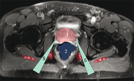
The Biopsy of the Prostate under MR-Guidance is typically carried out according to the High-Resolution MRI-Scan. Given Local Anesthesia, two Puncture needles transgluteal [5] (from behind by the Gluteal Muscles) in the Peripheral Zones of the Prostate [1] under MRI Control Ingested. Such access the Prostate Puncture can be carried out without damaging the Intestine [2] or the bladder.

After Final Position Control of the Cannulas before the Suspecten areas then takes multiple Biopsy using a Biopsy Punch. The maximum Duration of Treatment is 30-45 minutes. In order to avoid the occurrence of pain during the Procedure, there is a superficial and a deep Local Anesthesia.
After the Extraction of the tissue samples and final Biopsy, the removal of the Puncture Needles Occurs. Depending on the Course of Treatment, a CT-Scan is performed to detect possible Complication. After two hours of time spent monitoring and ambulatory Treatment is completed.

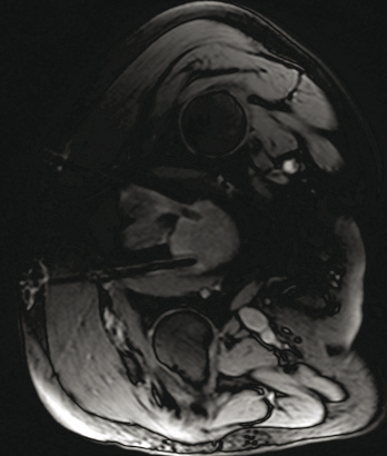
Representation of the Inserted needles into the Prostate gland (arrow) with positioning the bottom of tube before the suspicious Lesion shown in the MRI-Scan. MRI can be done to a safe Position control of the Puncture Instruments.

