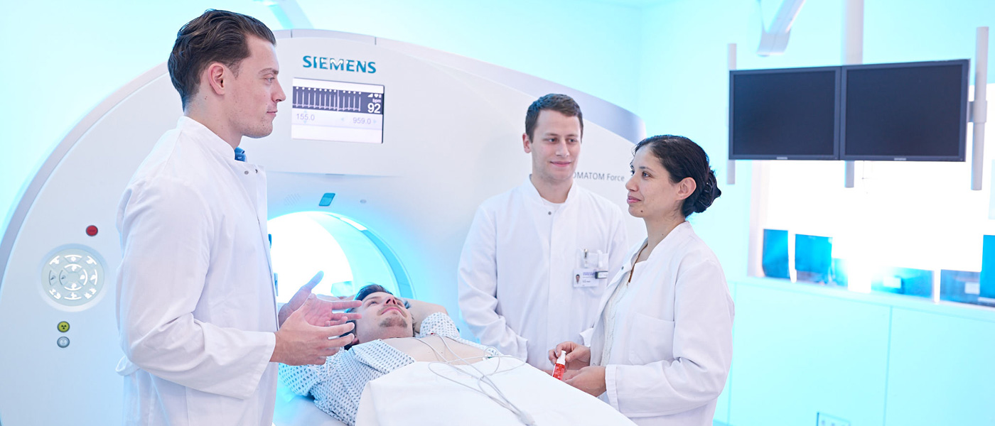
Diagnostics in the Computed Tomography
Magnet resonance imaging allows one to produce cross-sectional images of human body without the use of radiation. Radiowaves produced within a spacious magnetic tube causes reactions within the body which is received as signal by coils placed on the examining region. These signals are converted into images by computer.
An MRI examination usually takes about 20 to 40 minutes. The patient is placed supine in a 60 cm broad (diameter) and 160 long tube. In cases of claustrophobic patients examination can be performed under mild sedation.
During the examination the patient hears loud banging-type of noise which is produced by activation of electromagnet (Radiofrequency) and unfortunately cannot be avoided. To dampen the noise the patients are provided with a head phone or ear plugs. It is very important that the patient lies still during the examination. Even minimal movements can lead to decrease in image quality. For examinations of the abdomen and thorax one receives breathing commands and must hold breath for short periods (10 to 20 seconds). Majority of examinations are performed with a special MRI-contrast medial (gadolinium) in order to better differentiate the soft tissues.
For the examination it is also important that the patient removes all metal elements from his/her body (e.g. spectacles, watch, jeweleries, hearing aids, tooth-implantations etc.). Also small zip or clip in brassiere can lead to disturbances in the image.
Important: if you have a cardiac pace maker you should not in any case enter the examination room!
Procedure and preparation before a CT scan
Before the CT examination, an explanatory talk will take place in which you will be informed about possible complications. The doctor in charge will determine the individual examination procedure in consultation with you; you will decide with the radiologist whether a contrast medium will be administered for this purpose. A CT examination usually only takes a few minutes. You will usually lie on your back. The CT scanners at our institute have a very wide opening, so there is no reason for claustrophobia.
You must lie still during the examination. Even small movements can disturb the images. For examinations of the abdomen and chest area, you may have to hold your breath for a short time (10-20 seconds).
If you have a known allergy to contrast media, hyperthyroidism or kidney disease, the radiologist will make a decision together with you after carefully weighing up the risks and benefits.
If you suffer from diabetes and are being treated with metformin medication, you should stop taking this medication 2 days before the examination after consulting your family doctor.
Contraindications
There are no absolute contraindications for the performance of a CT examination. However, pregnant women should only be examined in exceptional cases due to the risk to the child from X-rays.
What you should bring to the examination
Reports and images of previous examinations are very helpful for planning and evaluating the computer tomography.
Please bring these with you if they were not performed at our institute.
For a CT examination, you should bring current laboratory values with you that provide information about your kidney function:
- Creatinine value
- Glomerular filtration rate (GFR)
- TSH (thyroid).
These should not be older than 14 days. For this purpose, you can have your blood drawn by your referring doctor or family doctor. Please bring the results of the laboratory test with you.
