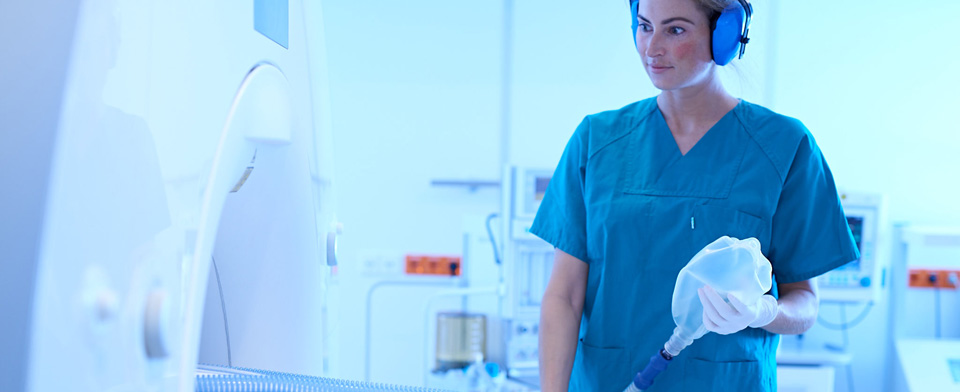
MR Mammography
Dear Patients
On the following pages we would like to inform you about MR-Mammography done on a daily basis at our institution.
History:
The MRI examination of the breast tissue began with the introduction of MR-contrast media in the free market in 1988 although scientific studies had already begun. Mrs. Heywang-Köbrunner played a major role in evaluation of MRI for diagnosis of breast carcinomas. In 1986 she published the first results [1] of application of contrast enhanced MRI in the field of breast diagnosis. The technical problems at the beginning of MR-era was firstly the low temporal resolution of the machines at that time and secondly the unavailability of breast compatible coil technology. Hence initially only static examination could be performed and only of one breast. The appropriate coil for simultaneous examination of both the breasts was developed several years later. Since the last few years simultaneous examination of both breast with dynamic sequences before and after contrast media application has become obligatory.
Besides, since the mid 90s MR-guided biopsy or MR-guided preoperative marking of uncertain breast lesions is being performed at many centres including ours. This way the high sensitivity of the procedure can be used just like with sonography for histological examination.
Technique
For the MRI examination of human body, magnetic field of about 15000 times (depending on machine) more that the earth's magnetic field is used. With the help of complex algorithm (Fourrier transformation) two dimensional images are created just like in computer tomography. The MRI examination of the breast requires a special technique for a high spatial and temporal resolution. The present standard technique consists of a dynamic simultaneous examination of both breasts with fat suppressed high resolution T1 weighted 3D sequences before and after contrast application in axial and coronal slices.
The main objective for contrast-dynamic is a spatial resolution of about 90 seconds per single sequence with a slice thickness of maximum 3 mm. In the course of the dynamic examination at least 5 measurements are performed after application of contrast media - with the most modern MR machines it is possible to obtain images of 1 mm slice thickness with good temporal resolution.
Additionally T2 weighted axial likewise T1 weighted coronal sequences are obtained. At the end of the examination the dynamic sequences after contrast media application are subtracted from the non-enhanced sequences and 3D-MIP (maximum intensity projection) are created. Suspected or pathological lesions with early arterial enhancement are evaluation using a special software
which analyses the signal intensity in relation to time.
For MR-guided biopsy in addition to a special coil which allows a lateral entry to the breast, a special software is required which calculates the co-ordination to the pathological lesion is required. Besides, special materials for biopsy/marking is required because standard biopsy instruments are not MRI compatible (could be pulled or lead to artifacts) and makes the exact localisation of the instrument impossible. The MRI compatible instruments are made of titanium and are readily available in the market.
Method:
The patient is firstly explained about the procedure and a consent is obtained. The patient is explained that the examination is harmless just like ultrasonography without any radiation, that metals should not be taken into the examination room, that a loud banging noise is heard during the examination, that the examination lasts about 30 minutes and that she must lie still without movement during the examination and the applied contrast media is very well tolerated. In cases of known allergy to contrast media, a premedication can undertaken to prevent the allergy. There is no fear of interaction with thyroid hormone induced metabolism. Serum creatinine level is required to rule out any renal insufficiency (a glomerular filtration rate of < 30 is contraindicated). An absolute contraindication is cardiac pacemaker - patients with pace maker should not enter the examination room. Also aneurysma clips are contraindicated. The modern cardiac valve prosthesis and port catheter system are no more considered an contraindication but in some cases the manufacturer must be questioned about the make of the prosthesis.
A peripheral i.v canula is placed and connected to the contrast media injector. The patient is then placed prone with each breast placed in a coil which is built in a support system.
Indications:
Because MRI examination is a complex and most expensive imaging modality, the indications should be carefully made taking in view the advantages of the procedure. The main indications for MRI examination of the breast are (in accordance to the recent literatures):
- 1. To rule out relapse of a breast carcinoma or verification of uncertain sonography and mammography finding.
- 2. In women with breast implantation due to cosmetic or oncological reasons to rule out a breast carcinoma or defect in the implantation.
- 3. Patient with metastases with unknown primary (CUP-Syndrome).
- 4. Patient with confirmed breast carcinoma and suspicion of multifocal/concentric or contralateral breast carcinoma.
- 5. Early diagnosis in women with gene mutation (as suggested by Deutschen Krebshilfe BRCA ½-Project - "German Cancer Help Organisation").
Sinful indications in special cases:
- 1. Women with breast which cannot be evaluated with sonography or mammography.
- 2. Women with pathological mammography or sonography finding without the possibility of a biopsy due to small size or non localisation in "second mammography view".
- 3. In women under neoadjuvant chemotherapy for preoperative evaluation of the size, morphology and localisation and in some cases relapses.
- 4. In women with preoperative marking in cases of clinical/sonographical/mammographical complete remission.
Literature:
- Heywang SH, Hahn D, Schmidt H, Krischke I, Eiermann W, Bassermann R, Lissner J MR imaging of the breast using gadolinium-DTPA. J Comput Assist Tomogr. 1986 Mar-Apr; 10(2): 199-204
- El Yousef, Duchesneau EH, Alfidi RJ, Haaga JR, Bryan PJ, Lipunk JP. Magnetic resonance of the breast. Work in progress. Radiology 1984 Mar; 150(3): 761-6
- Heywang SH, Hahn D, Eiermann W, Krischke I, Lissner J. Nuclear magnetic resonance tomography in breast cancer diagnosis- present status and future outlook. Digitale Bilddiagn. 1985 Sep; 5(3): 107-11
- Kaiser W, Zeitler E. Nuclear magnetic resonance tomography of the breast: diagnosis, differential diagnosis, problems and possible solutions II: Diagnosis Rofo 1986 May; 144(5): 572-9
- Kuhl CK, Mielcareck P, Klaschik S, Leutner C, Wardelmann E, Gieseke J, Schild H. Dynamic breast MR imaging: Are signal intensity time course data useful for differential diagnosis of enhancing lesions? Radiology 1999; 211:101-110
- Kuhl CK, Schild H. Dynamic image interpretation of MRI of the breast J Magn Reson Imaging 2000; 12: 965-974
- Mueller-Schimpfle M, Stoll p, Stern W, Kurz S, Dammann F, Claussen CD. Do mammography, sonography and MR Mammography have diagnostic benefit compared with mamography and sonography? Am J Roentgenol 1997; 168(5): 1323-9
- Kuhl CK MRI of breast tumors. Eur. Radiol. 2000 10, 46-58
- Fischer U, Kopka l, Grabbe E. Breast Carcinoma: Effect of preoperative Contrast-enhanced MR-Imaging on the therapeutic Approach. Radiology 1999. 213: 881-888
- Baum F, Fischer U, Vosshenrich R, Grabbe E. Classification of hypervascularized lesions in CE MR imaging of the breast. Eur Radiol. 2002 May; 12(5): 1087-92
- Heywang-Kobrunner SH, Bick U, Bradley WG Jr, Bone B, Casselman J, Coulthard A, Fischer U, Muller-Schimpfle M, Oellinger H, Patt R, Teubner J, Friedrich M, Newstead G, Holland R, Schauer A, Sickles EA, Tabar L, Waisman J, Wernecke KD International investigation of breast MRI: results of a multicentre study (11 sites) concerning diagnostic parameters for contrast-enhanced MRI based on 519 histopathologically correlated lesions Eur Radiol. 2001;11(4):531-46
- Hata T, Takahashi H, Watanabe K, Takahashi M, Taguchi K, Itoh T, Todo s. Magnetic resonance imaging for preoperative evaluation of breast cancer: A comparative study with mammography and ultrasonography. J Am Coll Surg. 2004 Feb; 198(2): 190-7
- Kuhl CK MRI of breast tumors. Eur. Radiol. 2000 10, 46-58
- Mueller-Schimpfle M, Ohmenhauser K, Stoll P, Dietz K, Claussen CD. Menstrual cycle and age: Influence on parenchymal contrast medium enhancement in MR-imaging of the breast. Radiology 1997 Apr; 203(1): 145-9
- Betsch A, Arndt E, Stern W, Wallwiener D, Claussen CD, Müller-Schimpfle M. Können Verlaufskontrollen die diagnostischen Sicherheit der MR-Mammographie erhöhen? Eine retrospektive Analyse MR-mammographischer Verlaufsuntersuchungen. Rofo 2001 Nov; 24-30
- Knopp MV, Bourne MW, Sardanelli F, Wasser MN, Bonomo L, Boetes C, Muller-Schimpfle M, Hall-Craggs MA, Hamm B, Orlacchio A, Bartolozzi C, Kessler M, Fischer U, Schneider G, Oudkerk M, Teh WL, Gehl HB, Salerio I, Pirovano G, La Noce A, Kirchin MA, Spinazzi A. Gadobenate dimeglumine-enhanced MRI of the breast: analysis of dose response and comparison with gadopentetate dimeglumine. AJR Am J Roentgenol. 2003 Sep;181(3):663-76.
- Mueller-Schimpfle M, Noack F, Oettling G, Haug G, Kienzler D, Gepp M, Dietz K, Claussen CD. Influence of histopathologic factors on dynamic MR mammography. Rofo 2000 Nov; 172(11): 894-900
