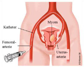
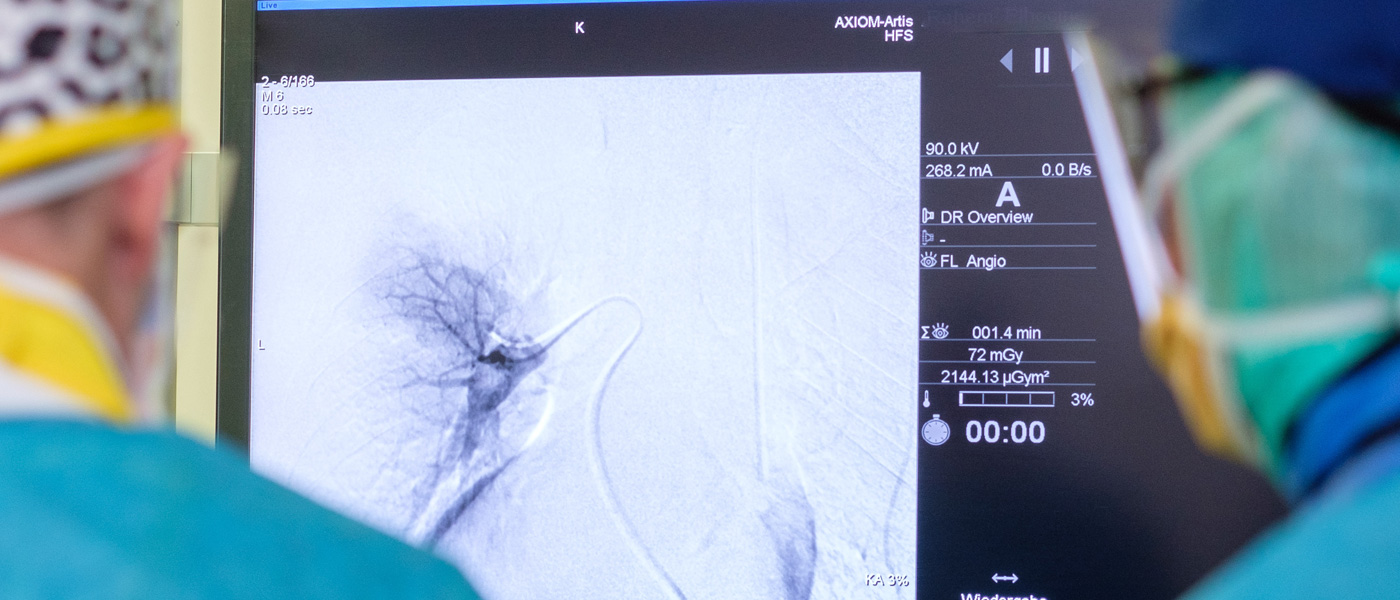
After local anaesthesia of the groin region, a special needle is used to puncture the femoral artery of the leg. A "lock" (a guide tube) is then inserted into the artery. A catheter (a thin flexible plastic tube) is then inserted through the sheath. Under angiographic visual control and contrast injections, the catheter is inserted into the uterine artery and finally into the vessels supplying the fibroid. The patient does not feel anything from the placement of the catheter. Only during the contrast medium injections does a feeling of warmth occur, which disappears within a few seconds. The administration of contrast medium makes the vessels visible in the angiography and serves to locate the myoma vessels.
Myoma embolisation
Dear Patients,
On the following pages we would like to inform you about the minimally invasive therapy procedure of myoma embolisation offered at our institute. With this procedure, the blood supply to the fibroids is interrupted by injecting tiny particles. As a result of the interrupted blood supply, the myoma nodes sclerose. The great advantage is that the uterus remains completely intact.

What are fibroids?
Myomas are benign tumours that form in the muscular wall of the uterus. They are the most common female abdominal tumours. The frequency is 35 - 50% of all women aged 35 - 50 years. The exact circumstances that contribute to the development and growth of fibroids are not all known.
The following factors have a favourable effect:
- Family history
- Obesity
- Childlessness
There are three types of fibroids, depending on their location, they have different names:
Intramural - within the muscle layer, usually lead to increased menstrual bleeding;
Submucous - below the endometrium;
Subserous - outside the uterine wall, can become very large without treatment;
Course of treatment
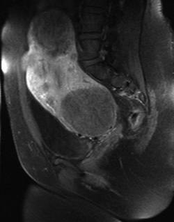
The treatment is performed entirely on an outpatient basis. You must present yourself at our institute the day before the procedure. Please bring all your medical reports and a current laboratory report with you to the appointment. During your stay, a magnetic resonance tomography will be performed to assess the location, number, size and blood flow of the fibroids. After the magnetic resonance imaging, the radiologist in charge will explain the procedure in detail.
On the treatment day
Some preparations will be made before the procedure. You will be given an indwelling cannula for an IV.
The procedure is performed by specially trained radiologists.
Procedure of myoma embolisation


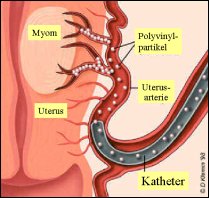
After correct placement of the catheter, the actual embolisation is performed. Under angiographic control, the embolisation particles are injected into the myoma vessels, ensuring that the blood supply and nutrition to the myoma is permanently cut off. The particles used for embolisation have a special size (500 to 900 µm) adapted for this purpose. This is chosen so that they mainly reach the fibroids and not the healthy uterine tissue.
After the myoma embolisation is completed, the catheter is removed from the inguinal artery and a pressure bandage is applied to the patient, which remains in place for 4-6 hours after the procedure. Afterwards, the patient should stay in bed for 6 hours. During this time, the patient's blood pressure and pulse are checked regularly and pain medication is administered if necessary.
Download further information:
Technical papers and scientific studies on myoma embolisation

