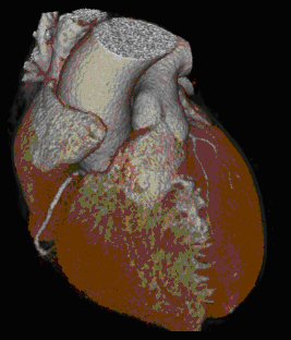
3D representation of Heart
Heart and Coronary Arteries Evaluation in Multislice Spiral Computed Tomography
In Institut for diagnostic and Interventionelle radiology of the Johann Wolfgang Goethe University clinic Frankfurt (you: Professor Dr. T. Vogl) are accomplished since end of 1999 not invasive investigations of the heart wreath/ring containers by means of multi-layer spiral CT. Until end of 2003 about 2100 patients became examined, approx. 500 investigations/year correspond. The photographs take place with the help of 16 lines a spiral CT's, which makes a simultaneous acquisition of 16 x for 0,75 mm possible of layers per 180° tube circulation. This corresponds to at present highest possible local dissolution and permits the evaluation of structures large up to 0,8 mm. The investigation is also accomplished lying on the back on the chest cork glued on electrodes. An electrocardiogram signal noted digitally during the admission makes following the investigation a reconstruction of transversals for sectional views possible by hearts in quasi the “frozen condition”. Thus artifacts will know the Koronararterien by the heart movement reduced and better to be defined. The duration of the investigation amounts to about 10 minutes, depending upon question varied thereby the number of CT-photographs. Thus for instance with the Kalkscoring in the context of a preventive medical examination a native CT-investigation is only needed, made during to the coronary angiography, bypass representation as well as with functional and pathomorphological questions additionally a contrast-centralstrengthened admission. For this during the second CT-investigation over a venous Verweilkanüle approx. 90 ml iodhaltiges contrast means is appliziert. Contraindications are a well-known heavy contrast central allergy - with lighter forms such as skin itching or - turning red can by simple medicamentous preparation possible symptoms in the apron be already avoided - as well as the income certain oral Antidiabetika (Metformin) in connection with a kidney insufficiency. Contrast means may not be given likewise with thyroid malfunctions, tumors of the thyroid as well as a planned radio iodine therapy. During each admission the patient must stop approx. 20 s air. Altogether different questions can be answered with the heart CT, which are in the following in detail represented.
Coronary Calcium Screening
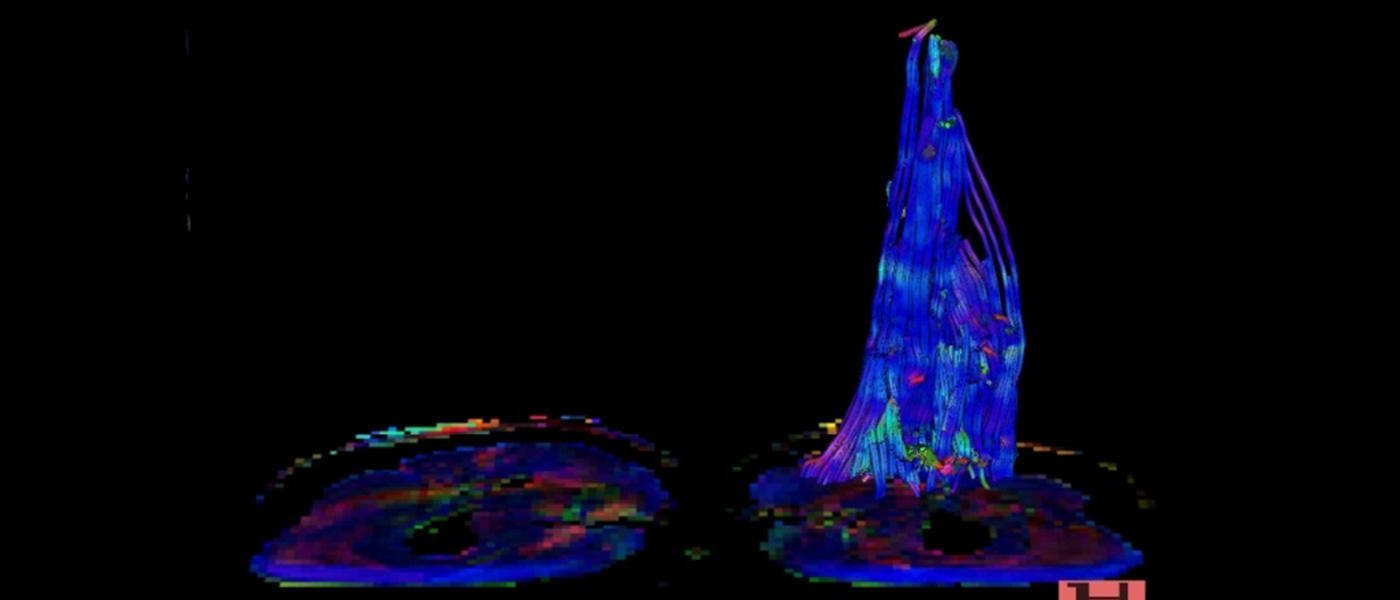
Coronary Calcium Screening refers to the Detection and Quantification of Coronary Artery Calcification. Similar studies have been carried out only with the Electron-beam CT (EBCT). Numerous Studies andSstudies of the past have shown that Coronary Calcifications have a high predictive value for the Probability of "Coronary" events. This Method is recommended especially in asymptomatic Patients with risk factors such. As Diabetes Mellitus, Hypertension, Family History and Hypercholesterolemia. If a pronounced Coronary Atherosclerosis detected in these Patients, further measures to modify the risk profile must be taken. The Result of these preventive Measures can then be monitored in more detail by way of follow-up of Coronary Calcification. In Addition, Patients with Atypical Heart Problems provide the simple exclusion or detection of Coronary Calcium groundbreaking Differential-Diagnostic Information. Technically, the Coronary Calcium Screening is performed without administration of Contrast Agent. This is an extremely gentle for the Patient clinically and easy to implement Method. The calcified Plaques are usually determined semiquantitatively according to the Agatston score. This Score takes into account all Areas with density Values above 130 HU in the Area of the Coronary Arteries. The Sum of all Values is the total Score Value and allows immediate Risk Assessment (Figure 1). For Values <10 the Risk of Coronary Heart disease is low, with Values <100 there is a high probability of Coronary Artery disease but without higher-grade Stenoses, with Values <400 are higher-grade Stenoses at least possible and likely at Values> 400. Definitively, this can be clarified, however, only in Vonjunction with Clinical findings and Contrast-Enhanced CTA.
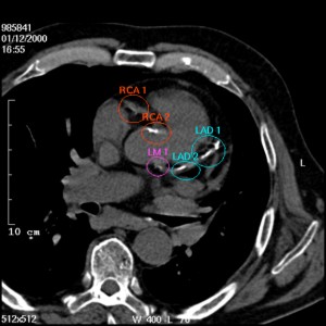
Detailed illustration of Coronary calcification in the Area right Coronary Artery (RCA), left Main Stem (LM) and left anterior descending Artery (LAD). Recording natively, prospective ECG triggering.
Cardiac CT Examinations
Other issues in Cardiac CT include the Presentation of Cardiac Tumors, the Assessment of Coronary Bypasses, Heart Valve Morphology and Function as well as answering Questions such as Functional Ejection Response,Eend-Systolic Ventricular Volume end-systolic or diastolic ventricular volume, post-ischemic post-ischemic Scarring or Wall Motion disturbances. Although this is usually the MRI Method of choice, but this must be decided individually from Patient to Patient.
CT Coronary Angiography
Calcification of the Coronary Arteries are an expression of advanced Atherosclerosis. The Quantification of Coronary Calcium can reveal the extent of Coronary Atherosclerosis. Lipid rich Plaques, often the real cause ischemia relevant vascular constrictions are, on the other hand, the Determination of Coronary Calcifications By not alone prove. So far, these Plaques were only indirectly Diagnosed with Cathetere Angiography and IVUS. With the new Multislice Spiral CT it is now possible Coronary Vessels up to a Diameter of about 1.0 mm reproducibly Display. In Addition to a very good axial and Multiplanar Imaging of the Vascular Course (Fig. 2) can be generated from the acquired Data Sets by Volume rendering Techniques also very nice 3-D Reconstructions.
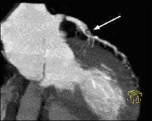
Fig.2 multiplanar reformation of the left coronary artery. Very nice view of the individual Septaläste and a 70% stenosis in the proximal vessel segment (white arrow).
Our previous Clinical Experience in 250 Patients, in comparison with the Cardiac Catheterization good agreement. However, the Indication for Cardiac CT should be done only in close Cooperation with the attending Cardiologist / Family Doctor and after previous careful clinical Examination.
Team:
Division of Experimental Imaging (DEI)
The Division of Experimental Imaging (DEI) at the Department of Diagnostic and Interventional Radiology at the Goethe University Hospital in Frankfurt, Germany is a research division focused on developing new imaging acquisition and post-processing techniques to ultimately improve imaging in clinical routine for our patients. Read more about us on our website...