Retikuloendothelial Contrast Media
AMI-25 (Endorem®), SHU-555 A (Resovist®)
Iron oxide enhanced MRI of focal and diffuse liver diseases
Optimization of Contrast Agents in MRI to improve the Evaluation of Liver Disease
In the last few years there has been a substantial increase in preooperative and imaging diagnostics with the modern advancement in oncological liver surgery and therapy of focal liver tumours. For this purpose in addition to standard methods like sonography, computer tomography and MRI there is also the new organ specific contrast enhanced MRI. The organ specific contrast allows improved diagnosis in terms of detection and localisation of liver lesions and also in terms of characterisation and diagnosis of the parenchyma.
Methods
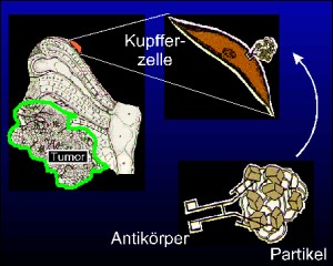
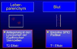
The iron oxide group belongs to the liver specific MRI contrast media which specially has an effect on the T2 relaxation time. The super paramagnetic iron oxide (SPIO) AMI-25 (Endorem®) is presently available for clinical use. A further smaller SPIO - SHU-555 A (Resovist®) is awaiting approval for clinical use. It is composed of iron-oxide crystals (Resovist®)enveloped by dextran (Endorem®) likewise Carboxydextran with an mean diameter of 150 nm likewise 58 nm. With a half life time of 8-10 min about 80% of the injected iron oxide particles are absorbed by the reticuloendothelial system (RES) of the liver through the Kupfer cells and about 12% are absorbed the RES of Spleen. The negative contrast enhancement is specially prominent in the proton density and T2 weighted sequences. A discrete T1 effect can be seen due to increase in T1 relaxation time.
Endoderm is given in dose of 15 µmol Fe/kg body weight through a filter canula in an infusion over a duration of 30 minutes. The application in 100 ml of 5% Glucose is done in two phases (2ml/min. and 4ml/min). Resovist in given in two different doses depending on body weight. It can be given as a bolus undiluted through a filter canula which allows dynamic studies of tumour vascularity and liver parenchyma.
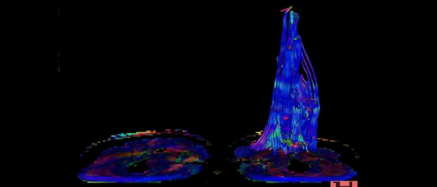
Effect of SPIO in MR-Imaging
Quantitative and Qualitative Criteria
The specific contrast enhancement of liver lesions is examined with non-enhanced and enhanced sequences. The loss of signal intensity can be used a useful criteria for pathological signal changes. Furthermore, other criteria like homogenity and form of lesions, the number of affected liver segments, number and size of lesions are interpreted. Presence of central changes like necrosis, scar and capsule are evaluated. The liver parenchyma is examined for changes like fibrosis, regenerative nodules, hypertrophy of liver segments and structure of surface of liver.
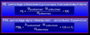
Formula for percentile contrast enhancement and loss of signal.
Results / Discussion
A total of 900 patients were examined in a 1.5 Tesla scanner (Siemens 63 SP; Philips ACS-NT) from May 1992 to December 1998. The sequence protocols were proton-density, T1w and T2w spin echo and gradient echo sequences before and after intravenous application of liver specific contrast media "Endorem".
Histopathology (liver resection, liver transplantation, fine-needle or punch biopsy) and clinical follow-up showed diagnosis of malignant (HCC, CCC, liver metastases, etc.) and benign liver lesions (FNH, adenoma, haemangioma, etc.).
For malignant tumors with regards to therapy relevant lesions iron-oxide enhanced MRI showed a sensitivity of 93.8% and a specificity of 91.5%, for benign lesions a sensitivity of 95.4% and a specificity of 93.9%.
In the group consisting of transplanted patients, the iron-oxide enhanced MRI with regards to diagnosis of HCC showed a sensitivity of 91.9% and a specificity of 86.7% in comparison to the gold standard. In comparison to preceding diagnosis (non-enhanced MRI / contrast enhanced CT) with gold standard technique, the contrast enhanced MRI showed a statistically significant better detection rate of malignant liver lesions (HCC and metastases) (p<0.05). The use of iron-oxide enhanced MRI brought a statistically significant changes in therapy decision in patients with HCC (p<0.05).
In a prospective phase II/III of the study, 82 patients with known liver tumors were examined with a 1.5 Tesla scanner (Siemens 63 SP; Philips ACS-NT). The sequence protocol included proton density, T2w, T1w SE and GRE sequences before and after intravenous application of superparamagnetic contrast media "Resovist".
The histopathological diagnosis showed malignant (n=49), borderline (n=2) and benign (n=9) liver lesions. The dynamic imaging after contrast application showed a significant drop-out phenomena in patients with highly differentiated HCC nodules: a rapid signal loss in the perfusion phase 40-90 seconds after contrast application, followed by a slow increase in signal intensity. The undifferentiated HCC nodules showed no loss of signal. The patients with FNH showed a significant signal loss beginning 30 seconds after contrast application followed by a even more loss of signal later.
Liver metastases from carcinoma of colon
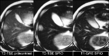
Hyperintense liver metastases in T2 weighted sequence. No evidence of signal loss in iron-oxide enhanced sequence. Conspicuous ring enhancement in iron-oxide enhanced T1 weighted sequence.
HCC in cirrhotic Hepatitis C
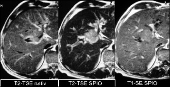
Evidence of hyperintense mass lesion in T2 weight sequence central in liver segment 8. No evidence of signal loss in iron-oxide enhanced sequence. Almost isointense signal in the T1w. Better definition of vascular structures.
Focal Nodular Hyperplasia
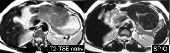
Evidence of hyperintense mass lesion in leftliver lobe in T2w sequence precontrast. Documentation of significant signal loss after application of iron-oxide contrast media.
Discussion
As a whole iron-oxide enhanced MRI showed a better detection rate and diffferentiation of focal liver lesions in comparison to conventional diagnostic methods. It is especially superior in patients with liver cirrhosis. This is an effective tool for detection and characterisation of malignant versus benign liver tumors with regards to therapy decision.
The dynamic iron-oxide enhanced MRI allows better evaluation of vascularity of the tumor and blood pool.