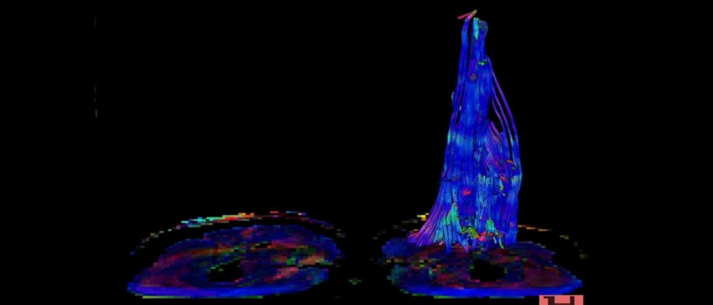
MRI Visualisation of HCC
Contrast-Enhanced MRI of Focal Liver Lesions
Optimization of Contrast Agents in MRI for better Evaluation of Liver Lesions
Introduction
In the last few years the diagnostic efficiency of MR-imaging of abdominal diseases has significantly increased with introduction of faster sequence protocols. However non-enhanced MRI leaves behind many questions with regards to detection and characterisation of focal liver lesions. With the help of liver specific contrast media most of these questions can be answered with regards to tissue specificity or hepatocyte function. Contrast medium such as tissue-specific contrast media, iron-oxide particles and also hepatobiliary contrast media belong to this group.
Methods
Three paramagnetic hepatobiliary contrast medium are presently available for clinical studies [Gd-EOB-DTPA (Eovist, Schering AG Berlin)] - likewise for clinical use [Mn-DPDP (Teslascan, Nycomed Imaging AS); Gd-BOPTA (MultiHance, Bracco-Byk-Gulden)]. Eovist and MultiHance are derivatives of Gd-DTPA where a carboxyl group is substitute by lipophilic side chain. About 50% (Eovist) and 2-4% (MultiHance) of injected dose is eliminated through the liver. The rest is eliminated through the kidneys. The presently suggested dose (phase 3) of Eovist is 25 µmol/kg of body weight. The suggested dose of MultiHance in clinical use is 0.05 mmol/kg of body weight. Teslascan contains DPDP-binded Mn which is slowly separated from DPDP in the plasma and reaches the liver cells through a selective transport mechanism. Free manganese are eliminated via the liver and kidney and also partly through pancreas and gastric mucosa. Its applications follows a short time infusion at the rate of 2-3 ml/ml in a dose of 0.5 ml/kg body weight (5µmol/kg body weight).
In intravenous application the Eovist and MultiHance spreads like extracellular contrast media and can be used for dynamic examinations. The hepatobiliary contrast media reaches the hepatocytes through an active transport mechanism. A late enhancement is achieved with Eovist after 10-20 minutes of application and after 40-90 minutes with MultiHance. Mn-DPDP leads to adequate positive hepatobiliary enhancement of liver parenchyma after completion of infusion.
Discussion
The use of hepatobiliary contrast media increases the detection rate of focal liver lesions in comparison to standard techniques. Dynamic examination protocol with application of Eovist and MultiHance also makes evaluation of the vascularity of the tumours. The characterisation of focal liver lesions with hepatobilary contrast media is similar to standard contrast media. The application of late scan protocols give additional informations about function of hepatocytes.
Results
In phase 2 of this study 57 patients with known liver lesions were prospectively examined in 1.5 Tesla scanner (Siemens). Aim of the study was to analyse the diagnostic accuracy in detection and differentiation of focal liver tumours with hepatobiliary contrast media Gd-EOB-DTPA in intraindividual comparison to standard contrast media Gd-DPTA.
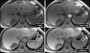
The sequence protocol consisted of static and dynamic T1-SE/GRE-sequences before, during and after intravenous application of Gd-EOB-DTPA (Eovist) in five different concentrations (3, 6, 12.5, 25 and 50 µmol/kg body weight). A Gd-DTPA (Magnevist: 0,1 mmo/kg body weight) enhanced MRI was performed within a week. Histopathology and clinical follow-up resulted in diagnosis of malignant liver tumours in 50 patients [HCC (n=22), CCC (n=2), metastases (n=26)] and benign liver lesions in 7 patients [FNH (n=4), haemangioma (n=3)]. Dynamic eovist-enhanced MRI documented a biphasic contrast enhancement in normal liver parenchyma (PE=154%), a lower enhancement in patients with liver cirrhosis (PE=78%). The HCC lesions showed increased peripheral enhancement as compared to metastases. An increased enhancement with eovist was seen in well-differentiated HCC nodules. The malignant lesions (HCC and metastases) document no contrast enhancement in the late phase sequences. The FNH lesions document strong contrast enhancement which increased even more in the late phase. The eovist-enhanced MRI allows better detection rate in comparison to non-enhanced and Gd-DTPA enhanced MRI (p<0.05). The percentile enhancement (PE) of malignant lesions was found to be statistically significant in comparison to benign lesions (p<0.05). As a whole the application of hepatobiliary contrast media eovist increases the detection rate of focal liver lesions in comparison to standard contrast media Gd-DTPA.
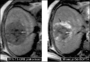
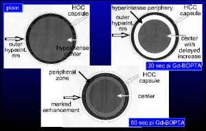
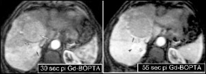
In the prospective phase 3 of our study 28 patients with histolopathological/clinical diagnosis of HCC were examined. A bolus injection of hepatobiliary contrast media Gd-BOPTA (MultiHance) was given in a concentration of 0.10 mmol/kg body weight. The sequence protocols included T1-SE/GRE sequences before, during and after intravenous application of Gd-BOPTA. A differentiation of highly differentiated and undifferentiated HCC nodules could be documented with dynamic imaging. Highly differentiated nodular HCC (< 3cm in size) showed a rapid increase in signal in the arterial phase with a solid blush. The larger nodular lesions showed double-ring sign: hyperintense rim precontrast, hypo and hyperintense rim postcontrast. The undifferentiated HCC nodules showed no blush in the dynamic imaging.
Dynamic Gd-BOPTA - enhanced MRI allows better differentiation of HCC nodules.
Gd-EOB-DTPA - Eovist
Dynamic MRI in Patients with Liver Metastases
Static MRI in Patients with Liver Metastases.
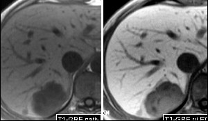
Fokal Nodular Hyperplasia
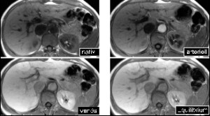
Highly Differentiated HCC
Undifferentiated HCC
MRI post contrast application in T2W
