Our Institute uses for Ultrasound Sonography Equipment of the latest Generation one with a frequency range of 2.5 MHz to 13 MHz, which are specially designed for the body to be examined Regions and Issues
Ultrasound (sonography)
Ultrasound diagnostics (sonography) is, along with computer tomography and magnetic resonance imaging, the most widely used imaging examination procedure in medicine. By emitting and receiving harmless ultrasound waves, this technique makes it possible to visualise most structures of the internal body. A gel is used for the examination because the sound waves cannot pass through air and bone.
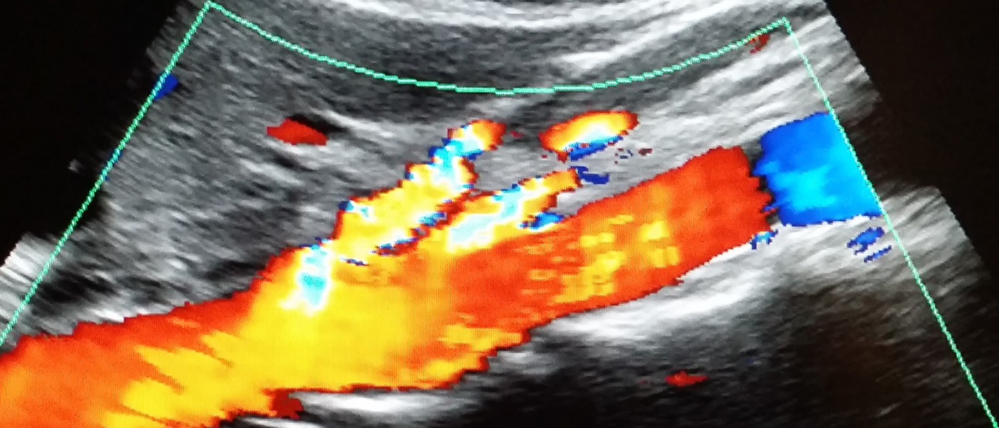
An ultrasound examination produces cross-sectional images. With the Doppler method, it is possible to visualise movement. With this procedure it is possible to assess the function of the heart muscle, the intestine and joints.
- Doppler examinations of the carotid artery, pelvic and leg veins, heart,...
- Lymph nodes
- Pancreas
- Liver
- Spleen
- Kidneys


Ultrasound of the kidneys

Doppler-Sonography of vessels
The doppler Sonography is a special ultrasonic investigation to measure blood velocity within the vessels (Arteries and Veins) and also to examine the vascularisation of tumors and lymph nodes.